Efficient Dissociation Protocol for Generation of Single Cell Suspension from Human Thyroid Tissue for Single Cell RNA Sequencing
Article information
Single cell RNA sequencing technology is a cutting-edge field that is currently in the spotlight for diverse research applications, such as the identification of rare cell populations or specific markers for specific small cell clusters. In particular, several methods have been suggested for the primary culture system for the ex vivo tissue structure manufactured from the thyroid tissue of a patient [1-3]; however, these are not suitable for single cell RNA sequencing as the manufacturing success rate is very low owing to the low cell viability. Here, we report an efficient dissociation protocol for generation of single cell suspensions from human thyroid tissue for single cell RNA sequencing. The present protocol method comprises the following steps: (1) transferring the separated thyroid tissue to a laboratory while immersed in a low-temperature storage solution; (2) mechanical digestion by chopping of the tissue in the culture dish and transferring it to a transfer container; (3) chemical digestion by reacting with a mixed solution of trypsin and collagenase type 4; and (4) removal of red blood cells and dead cells. The detailed methods are presented below.
1. Collect the biopsy sample and place in a 50 mL conical tube containing 5 mL of Hanks′ Balanced Salt solution (HBSS) (Gibco, Cat# 14025-076, Waltham, MA, USA) on ice and transfer to the laboratory within 15 minutes.
2. Discard the supernatant, add Roswell Park Memorial Institute (RPMI)-1640 (Welgene, Cat# LM011-01, Taipei, Taiwan) Fetal Bovine Serum (FBS) with Red Blood Cell (RBC) lysis buffer (Roche, Cat# 1181438-9001, Basel, Switzerland); then, wash and centrifuge at 4,000 rpm for 5 minutes at 4°C.
a. Hold the tissue sample with forceps and cut it using a scissor.
b. Continue cutting so that the tissue is minced into pieces until it can be moved using the bore tip.
3. Quickly transfer the minced biopsy tissue to a 15 mL tube.
4. Add enzyme digestion solution to the tube and mix well.
a. Prepare the digestion solution by mixing 0.125% trypsin (Sigma Aldrich, Cat# T1426, St. Louis, MO, USA), 1:300 unit/mL collagenase IV (Worthington, Cat# LS004188, Columbus, OH, USA), and 4 mL of RPMI-1640 (FBS free).
b. After the digestion solution is completely mixed, add it to the minced biopsy tissue.
5. Place the tube containing the biopsy tissue and enzyme digestion solution in a shaking incubator at 37°C and 100 rpm for 60 minutes.
6. Centrifuge at 4,000 rpm for 5 minutes at 4°C.
7. Discard the supernatant and add 1 mL of RPMI-1640 (FBS+; Gibco, Cat# 16000044) to the digested tissue.
8. Perform mechanical digestion using wide bore tips.
a. Pipet approximately 20 times through a 1 mL wide bore tip so that the tissue clumps in the solution are not visible to the naked eye.
b. Pipet approximately 50 times at multiple locations in the solution with a 200 μL wide bore tip.
9. Load the sample on a 70 μm pore cell strainer and add 500 μL of RPMI-1640 (FBS+).
Optional: If there is a lot of tissue above the strainer, add an additional 500 μL of RPMI-1640 (FBS+).
10. Centrifuge at 4,000 rpm for 5 minutes at 4°C.
11. Discard the supernatant and mix briefly with 1 mL of RBC lysis buffer (Roche, Cat# 11814389001).
a. Incubate the tube on ice for 10 minutes with tapping the tube every 2 minutes.
b. After 10 minutes, centrifuge at 4,000 rpm for 5 minutes at 4°C.
12. Discard the supernatant, add RPMI-1640 (FBS–) with RBC lysis buffer; then, wash and centrifuge at 4,000 rpm for 5 minutes at 4°C.
13. Discard the supernatant and re-suspend in 300 μL of RPMI-1640 (FBS–).
14. Place the sample on a 40 μm pore cell strainer and add 200 μL of RPMI-1640 (FBS+).
15. Remove dead cells
a. Remove dead cells using the microbeads in the Dead Cell Removal kit (Miltenyi Biotec, Catalogue number: 130-090-101, Gaithersburg, MD, USA).
Optional: If the sample contains cell clumps, strain the sample using 30 μm pre-separation filters to remove clumps before using the Dead Cell Removal kit.
b. Perform the experiment according to the protocol in the data sheet provided with the cell counter (LUNA-FX7™ Automated Cell Counter, Logos Biosystems, Anyang, USA).
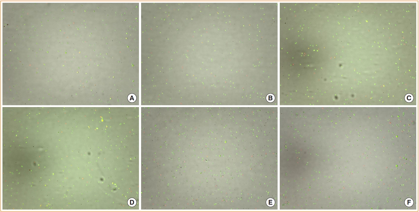
We successfully isolated single cell from normal thyroid tissue, differentiated thyroid cancer tissue, and anaplastic thyroid cancer tissue using the above protocol (Fig. 1). In conclusion, we developed the efficient dissociation protocol for generation of single cell suspensions from human normal thyroid tissue and thyroid tumor tissues for single cell based research.

Representative images of the cell counter (LUNA-FX7™ Automated Cell Counter, Logos Biosystems). (A) Ninety-three percent viability in the normal thyroid sample from the patient with papillary thyroid microcarinoma after the dissociation protocol (50×). (B) Eighty-six percent viability in the normal thyroid sample from the patient with adenomatous goiter after the dissociation protocol (50×). (C) Ninety-one percent viability in the tumor sample from the patient with papillary thyroid carcinoma after the dissociation protocol (50×). (D) Ninety-three percent viability from the tumor samples from the patient with follicular thyroid carcinoma after the dissociation protocol (50×). (E) Eighty-four percent viability from the tumor samples from the patient with anaplastic thyroid carcinoma after the dissociation protocol (50×). (F) Eighty-six percent viability from the tumor samples from the patient with anaplastic thyroid carcinoma after the dissociation protocol (50×).
This study was approved by the Institutional Research and Ethics Committee at Chungnam National University Hospital (CNUH- 2020-11-004-001). Written informed consent was obtained from all patients.
Notes
CONFLICTS OF INTEREST
No potential conflict of interest relevant to this article was reported. There was no potential conflict of interest with Macrogen Inc.
Acknowledgements
This research was supported by a grant from the Korea Health Technology R&D Project through the Korea Health Industry Development Institute, funded by the Ministry of Health & Welfare, Republic of Korea (grant number: HR20C0025) and the National Research Foundation of Korea (NRF) (grant number: 2021R1C1C1011183) to Yea Eun Kang.
