
Articles
- Page Path
- HOME > Endocrinol Metab > Volume 30(2); 2015 > Article
-
Case ReportPartial Androgen Insensitivity Syndrome Presenting with Gynecomastia
- Sung Won Lee, Dong Shin Kwak, In Sub Jung, Joo Hee Kwak, Jung Hwan Park, Sang Mo Hong, Chang Bum Lee, Yong Soo Park, Dong Sun Kim, Woong Hwan Choi, You Hern Ahn
-
Endocrinology and Metabolism 2015;30(2):226-230.
DOI: https://doi.org/10.3803/EnM.2015.30.2.226
Published online: June 30, 2015
Department of Internal Medicine, Hanyang University College of Medicine, Seoul, Korea.
- Corresponding author: You Hern Ahn. Department of Internal Medicine, Hanyang University College of Medicine, 222-1 Wangsimni-ro, Seongdong-gu, Seoul 133-792, Korea. Tel: +82-2-2290-9556, Fax: +82-2-2290-8231, ahnyh@hanyang.ac.kr
• Received: August 4, 2014 • Revised: September 1, 2014 • Accepted: September 3, 2014
Copyright © 2015 Korean Endocrine Society
This is an Open Access article distributed under the terms of the Creative Commons Attribution Non-Commercial License (http://creativecommons.org/licenses/by-nc/3.0/) which permits unrestricted non-commercial use, distribution, and reproduction in any medium, provided the original work is properly cited.
ABSTRACT
- Gynecomastia is a benign enlargement of the male breast caused by the proliferation of glandular breast tissue. Determining the various causes of gynecomastia such as physiological causes, drugs, systemic diseases, and endocrine disorders is important. Androgen insensitivity syndrome (AIS) is a rare endocrine disorder presenting with gynecomastia and is a disorder of male sexual differentiation caused by mutations within the androgen receptor gene. All individuals with AIS have the 46 XY karyotype, although AIS phenotypes can be classified as mild, partial or complete and can differ among both males and females including ambiguous genitalia or infertility in males. We experienced a case of partial AIS presenting with gynecomastia and identified the androgen receptor gene mutation.
- Gynecomastia is a benign enlargement of the male breast caused by proliferation of glandular breast tissue and is seen occasionally in clinical practice. The incidence and prevalence of gynecomastia are estimated at 3% to 40% and vary according to age. Physiologic gynecomastia is common and can be found in infants, adolescents and the elderly. Drugs and systemic diseases can cause pathologic gynecomastia. In young patients, determining the causes of gynecomastia such as testicular tumor, adrenocortical tumor, primary hypogonadism, Klinfelter's syndrome and rarely androgen insensitivity syndrome (AIS) is necessary.
- We experienced a case of partial AIS which presented with gynecomastia, in whom we identified an androgen receptor gene mutation. Herein, we report this case along with the relevant literature.
INTRODUCTION
- A 16-year-old male was referred and presented to our clinic with bilateral breast enlargement for 4 years (Fig. 1). Because of gynecomastia, he was withdrawn and avoided outdoor activities. He was born with hypospadias and underwent surgery at 1 year of age. The patient was 161 cm in height, 57.8 kg in weight (body mass index, 22.3 kg/m2), and his blood pressure was 120/80 mm Hg. His pubertal development was normal, but both breasts were enlarged (Tanner stage III). Pubic hairs were observed (Tanner stage V), the volume of both testes was 15 mL (Tanner stage IV), and a micropenis was noticed (Fig. 2).
- Complete blood count, biochemistry and urinalysis were normal. Endocrine results were as follows: testosterone level of 50.83 nmol/L (normal range, 9.7 to 27.8), dihydroepiandrosterone (DHEA) level of 15.03 nmol/L (normal range, 6.2 to 43.3), and DHEA-sulfate (DHEA-S) level of 5,120 nmol/L (normal range, 1,600 to 12,200), 17-hydroxyprogesterone level of 6.61 nmol/L (normal range, 1.88 to 10.94), lutenizing hormone (LH) level of 11.5 IU/L (normal range, 1.7 to 8.6), follicle stimulating hormone (FSH) level of 5.5 IU/L (normal range, 1.5 to 12.4), estradiol level of 0.191 mmol/L (normal range, 0.026 to 0.147), β-human chorionic gonadotropin (β-hCG) level of <0.1 IU/L (normal range, <2.6), prolactin of 21.7 pg/L (normal range, 4.0 to 15.2), free T4 level of 18.9 pmol/L (normal range, 11.9 to 21.9), and thyroid stimulating hormone level of 2.43 mIU/L (normal range, 0.27 to 4.2).
- The size of the right testis was 4.3×2.0×2.6 cm, and the left was 4.4×2.1×1.8 cm based on the ultrasound. Multiple microliths were present in both testes. The seminal vesicles and prostate glands were normal in size and echogenicity. Sellar magnetic resonance imaging and abdominal computed tomography (CT) showed no significant findings. His karyotype was male (46, XY).
- To determine the androgen receptor gene mutation, the androgen receptor gene was amplified using polymerase chain reaction (PCR) with exon-specific probes and sequenced. A somatic mosaicism (c.2128G>A, [p.Glu710Lys]) in exon 4 was found (Fig. 3).
- The patient underwent mammoplasty without complications. He was satisfied with the surgery results and was followed up in the outpatient clinic.
CASE REPORT
- Gynecomastia is defined histologically as a benign proliferation of the male breast's glandular tissue and clinically by the presence of a rubbery or firm mass extending concentrically from the nipples. Gynecomastia is common at any age from infancy to the elderly and the incidence and prevalence vary according to age [1].
- The pathophysiological process of gynecomastia involves an imbalance between free estrogen and free androgen actions in the breast tissue [2]. There are various causes of gynecomastia. Physiologic gynecomastia is the most common and is seen in infants, adolescents and the elderly. In our case, the patient was born with hypospadias and underwent surgery at 1 year of age and his testes were descended.
- Drugs are the second most common cause of gynecomastia (10% to 25% of all gynecomastia cases). Numerous drugs exert a mechanism related to that of exogenous estrogen, such as oral contraceptives, tamoxifen, and estrogen-containing creams. Additionally, other drugs have mechanisms related to antiandrogen therapies, such as bicalutamide, finasteride, and others with unclear mechanisms, such as tricyclic antidepressants, furosemide, digitalis, ketoconazole, and cimetidine. Our patient denied taking any drugs related to gynecomastia.
- Systemic diseases, such as hyperthyroidism, liver cirrhosis [3], chronic kidney disease [4], chronic pulmonary disease, and malignancy, can cause pathologic gynecomastia. In young patients, it is very important to exclude rare causes such as testicular tumors (Leydig-cell tumor or Sertoli-cell tumor), adrenocortical tumor, primary hypogonadism, Klinfelter's syndrome, and rarely AIS [5]. Additionally, in some cases of adult-onset gynecomastia, the etiology remains idiopathic [6]. In our case, systemic diseases such as hyperthyroidism, liver cirrhosis, renal insufficiency, and pulmonary disease were excluded based on the physical examination, blood tests, and radiological findings. Endocrine tests showed undetectable β-hCG and normal 17-hydroxyprogesterone, DHEA, and DHEA-S levels, while abdominal CT with contrast enhancement and scrotal ultrasonography showed unremarkable findings. Therefore, testicular tumors and adrenocortical tumors were excluded. Because the levels of LH and estradiol were increased while the level of FSH was normal in the presence of a high testosterone level, we excluded primary hypergonadism and instead considered AIS as the cause of gynecomastia.
- A diagnosis of AIS requires the following laboratory findings: a 46, XY karyotype, normal or increased serum testosterone, normal conversion of testosterone to dihydrotestosterone, slightly increased LH due to absence of negative feedback [7], normal or slightly increased FSH and slightly increased estradiol due to excess testosterone production and peripheral aromatization to estrogen.
- The development of external genitalia during sex differentiation is determined by the action of androgens and androgen receptors. The androgen receptor is essential for androgen action, thus mutations in the androgen receptor gene can alter receptor function, which leads to AIS [8]. AIS can be subdivided into three broad phenotypes, complete, partial, and mild AIS.
- The typical presentation for complete AIS is a phenotypically normal female and either primary amenorrhea during adolescence or inguinal swellings in an infant. The patient undergoes breast development and a pubertal growth spurt at the appropriate age, but without menses. Pubic hair and axillary hair are usually absent or may be present in sparse amounts. Extragenital abnormalities are absent, but the vagina varies from a dimple in the perineum to normal length and is always blind-ending. The uterus, cervix, and proximal vagina are absent due to the action of antimüllerian hormone produced by Sertoli cells of the testes. Testes may or may not be inguinal.
- Depending on the degree of responsiveness of the external genitalia to androgens, the clinical presentation of partial AIS varies [9]. Partial AIS represents a wide range of undermasculinization from ambiguous genitalia to mild hypospadias. The various phenotypes include a micropenis, severe hypospadias and a bifid scrotum with potential presentation of gynecomastia, cryptorchism, or infertility. In another form, the external genitalia may be nearly normal female, except for clitoromegaly and/or posterior labial fusion. A grading scheme has been proposed by Quigley et al. [8] to describe the degree of masculinization in AIS patients.
- Mild AIS is the rarest form. There is no extragenital abnormality but it may present in males as infertility. It can also manifest as bulbar and spinal muscular atrophy (Kennedy's disease).
- Our patient had external genitalia of the male phenotype, thus complete AIS was excluded. And he had a medical history of hypospadia, micropenis, and gynecomastia. Because the patient had a history of ambiguous genitalia during childhood, mild AIS was also excluded. Therefore, his phenotype was compatible with that of partial AIS.
- The Androgen Insensitivity Syndrome Patient Database at the University of Cambridge reports that mutations are present in 95% of their complete AIS patients, but in only 25% of their partial AIS patients [10]. In Korea, a study analyzed androgen receptor mutations and found in six out of nine patients with complete AIS and one out of three patients with partial AIS [11].
- The androgen receptor is encoded by the androgen receptor gene located on the X-chromosome at Xq11-12 and consists of eight exons and seven introns that span ~90 kb of DNA. In 70% of cases, the mutations are germline mutations transmitted in an X-linked manner via the carrier mothers. The androgen receptor contains four different functional domains: an amino-terminal domain encoded by exon 1, which is a nonconserved region involved in transcriptional activation of target genes; a central DNA-binding domain encoded by exons 2 and 3, which contains two zinc finger motifs; a hinge region containing the nuclear targeting signal and a C-terminal ligand-binding domain encoded by exons 4 to 8, which also encompasses subdomains involved in dimerization and transcriptional activation processes. Most mutations of the androgen receptor gene are located within the ligand-binding domain. More than 800 mutations of the androgen receptor gene from different patients have been entered into the Cambridge database as of September, 2011 [10].
- In our case, to determine the androgen receptor gene mutation, direct PCR sequencing was performed. DNA was extracted from peripheral blood leukocytes and the eight exons and exon-intron boundaries of the androgen receptor gene were screened. The androgen receptor gene mutation was identified as a somatic mosaicism (c.2128G>A, [p.Glu710Lys]) in exon 4. This mutation was previously identified in a partial AIS patient in 2006 [12]. Additional androgen receptor gene mutation analyses for the family members of the patient were necessary. We attempted to persuade the family members to undergo genetic analysis for the androgen receptor gene, but they refused testing. Based on the identified androgen receptor gene mutation and characteristic phenotypes, a diagnosis of partial AIS was made.
- The patients with complete AIS have customary feminine appearance and are usually raised as females. Several studies noted that tumor risk increases with age, exceeding 30% for people in their fifties [13], indicating the necessity for gonadectomy and estrogen replacement after surgery. Their proper management and psychological support can give them a normal life, despite a major chromosomal discrepancy.
- Most infants with partial AIS are raised as males. Androgen supplementation may be necessary at puberty, but not always. Surgery is performed during the second to third year of life to repair hypospadias and bring undescended testes into the scrotum. Gynecomastia often occurs during adolescence and requires reduction mammoplasty. An infant with partial AIS who is designated a female will require genitoplasty and gonadectomy before puberty to avoid the risk of virilization. Estrogen replacement is needed to induce female puberty. The risk of germ cell tumors is higher in partial than complete AIS patients, with a suggested incidence of 15%, and is even higher if the testes are not scrotal in position [14]. Because their gender identity may be vague, psychological distress is more common in adults with partial AIS than in those with complete AIS [15].
- Our patient developed gynecomastia (Tanner stage III) at puberty. He did not complain of any decreased libido or difficulties in erection or ejaculation. However, the severity of gynecomastia made him withdrawn, and he avoided outdoor and social activities; work-up for gynecomastia resulted in a diagnosis of partial AIS. Subsequently, mammoplasty was performed. He was satisfied with the surgery results and was followed up in the outpatient clinic.
- Therefore, a male patient complaints of gynecomastia must be considered by physicians for physiological causes, drugs, systemic diseases such as liver cirrhosis, chronic kidney disease, hyperthyroidism and malignancy, as the common causes of gynecomastia. Particularly, if the patient is young, testicular tumors such as Leydig-cell or Sertoli-cell tumor, adrenocortical tumor, primary hypogonadism and Klinfelter's syndrome must be ruled out. If levels of testosterone, LH and FSH are normal or elevated, AIS should be considered.
DISCUSSION
-
CONFLICTS OF INTEREST: No potential conflict of interest relevant to this article was reported.
Article information
- 1. Eckman A, Dobs A. Drug-induced gynecomastia. Expert Opin Drug Saf 2008;7:691–702. ArticlePubMed
- 2. Mathur R, Braunstein GD. Gynecomastia: pathomechanisms and treatment strategies. Horm Res 1997;48:95–102. ArticlePubMed
- 3. Kee CS, Park DI, Lee OY, Han DS, Son JH, Yoon BC, Ahn YH, Ham JS, Lee MH, Park KN. Gynecomastia in patients with cirrhosis. Korean J Gastroenterol 1994;26:842–849.
- 4. Sung KJ, Lee MW, Choi JH, Koh JK, Park SK. The frequency of the cutaneous problems and the influence of hemodialysis in patients with chronic renal failure. Korean J Dermatol 1991;29:313–321.
- 5. Cho HJ, Yang S, Oh PS, Shin JH. A case of sertoli cell tumor presented with sexual precosity. J Korean Soc Pediatr Endocrinol 2004;9:86–90.
- 6. Braunstein GD. Clinical practice. Gynecomastia. N Engl J Med 2007;357:1229–1237. ArticlePubMed
- 7. Melo KF, Mendonca BB, Billerbeck AE, Costa EM, Inacio M, Silva FA, Leal AM, Latronico AC, Arnhold IJ. Clinical, hormonal, behavioral, and genetic characteristics of androgen insensitivity syndrome in a Brazilian cohort: five novel mutations in the androgen receptor gene. J Clin Endocrinol Metab 2003;88:3241–3250. ArticlePubMed
- 8. Quigley CA, De Bellis A, Marschke KB, el-Awady MK, Wilson EM, French FS. Androgen receptor defects: historical, clinical, and molecular perspectives. Endocr Rev 1995;16:271–321. ArticlePubMedPDF
- 9. Ahmed SF, Khwaja O, Hughes IA. The role of a clinical score in the assessment of ambiguous genitalia. BJU Int 2000;85:120–124. ArticlePubMed
- 10. Gottlieb B, Beitel LK, Nadarajah A, Paliouras M, Trifiro M. The androgen receptor gene mutations database: 2012 update. Hum Mutat 2012;33:887–894. ArticlePubMed
- 11. Park SY, Choi YM, Park SH, Yang SW, Ku SY, Kim SH, Kim SW, Paik JS, Yang DH, Choi DS, Kwon HC, Choi DH, Lee SH. Analysis of androgen receptor gene in Korean patients with androgen insensitivity syndrome. Korean J Obstet Gynecol 2001;44:655–662.
- 12. Georget V, Bourguet W, Lumbroso S, Makni S, Sultan C, Nicolas JC. Glutamic acid 709 substitutions highlight the importance of the interaction between androgen receptor helices H3 and H12 for androgen and antiandrogen actions. Mol Endocrinol 2006;20:724–734. ArticlePubMedPDF
- 13. Alvarez NR, Lee TM, Solorzano CC. Complete androgen insensitivity syndrome: the role of the endocrine surgeon. Am Surg 2005;71:241–243. ArticlePubMed
- 14. Cools M, Drop SL, Wolffenbuttel KP, Oosterhuis JW, Looijenga LH. Germ cell tumors in the intersex gonad: old paths, new directions, moving frontiers. Endocr Rev 2006;27:468–484. ArticlePubMedPDF
- 15. Bouvattier C, Mignot B, Lefevre H, Morel Y, Bougneres P. Impaired sexual activity in male adults with partial androgen insensitivity. J Clin Endocrinol Metab 2006;91:3310–3315. ArticlePubMedPDF
References


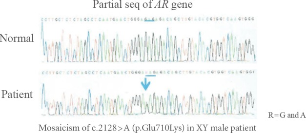
Figure & Data
References
Citations
Citations to this article as recorded by 
- A case of mild partial androgen insensitivity syndrome in a juvenile boy
Fen Wang, Shiying Shao, Wentao He, Shuhong Hu
Journal of International Medical Research.2024;[Epub] CrossRef - Clinical outcomes and genotype-phenotype correlations in patients with complete and partial androgen insensitivity syndromes
Nae-yun Lee, Ja Hye Kim, Ji-Hee Yoon, Soojin Hwang, Gu-Hwan Kim, Han-Wook Yoo, Jin-Ho Choi
Annals of Pediatric Endocrinology & Metabolism.2023; 28(3): 184. CrossRef - The Impact and Management of Gynaecomastia in Klinefelter Syndrome
Amr Abdel Raheem, Ahmed Said Zaghloul, Ahmed M. G. Sadek, Bilal Rayes, Tarek M. Abdel-Raheem
Frontiers in Reproductive Health.2021;[Epub] CrossRef - Identification of Potential Genes in Pathogenesis and Diagnostic Value Analysis of Partial Androgen Insensitivity Syndrome Using Bioinformatics Analysis
Yajie Peng, Hui Zhu, Bing Han, Yue Xu, Xuemeng Liu, Huaidong Song, Jie Qiao
Frontiers in Endocrinology.2021;[Epub] CrossRef - Adolescent Gynecomastia due to Minimal Androgen Resistance Syndrome: A Case Report and Literature Review
Aureliano Fiorini, Margherita Sepich, Margherita Pontrelli, Giorgio Sangriso, Mirna Cosci o Di Coscio, Marcella Lauletta, Fulvia Baldinotti, Diego Peroni, Maria Rosaria Ambrosio, Silvano Bertelloni
Sexual Development.2020; 14(1-6): 21. CrossRef - Endocrine and molecular investigations in a cohort of 25 adolescent males with prominent/persistent pubertal gynecomastia
F. Paris, L. Gaspari, F. Mbou, P. Philibert, F. Audran, Y. Morel, A. Biason‐Lauber, C. Sultan
Andrology.2016; 4(2): 263. CrossRef
 PubReader
 Cite
Cite


